Rapid 3D Brain Imaging Using Chaotic Laser Light: A Step-by-Step Protocol
By
Introduction
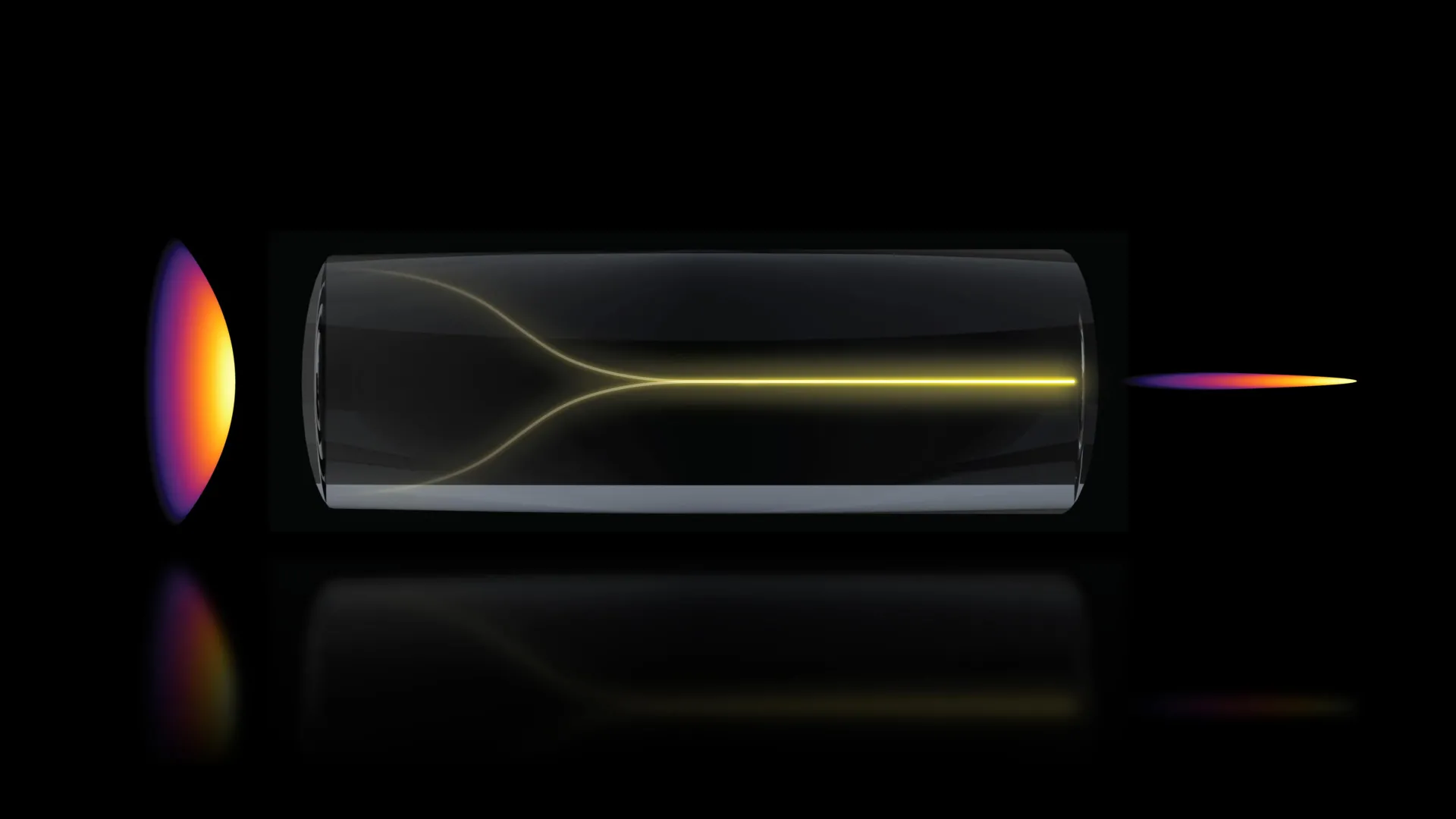
Imagine turning a chaotic laser beam—normally scattered and diffuse—into a highly focused pencil beam that slices through biological tissue. Scientists at MIT achieved exactly that, unlocking a revolutionary technique for imaging the blood-brain barrier (BBB) in 3D at speeds up to 25 times faster than conventional methods. This breakthrough allows researchers to watch in real time how drugs penetrate brain cells, dramatically accelerating the development of treatments for neurological diseases. In this guide, we will walk through the essential steps to replicate this groundbreaking imaging method, from setting up the laser source to capturing real-time drug dynamics.
What You Need
- High-power laser source (e.g., titanium-sapphire laser capable of producing chaotic emission).
- Optical components: lenses, mirrors, spatial light modulators, and a precision alignment stage.
- Chaos-inducing element (e.g., a saturable absorber or a feedback mirror placed at critical distance).
- Beam characterization tools: CCD camera, power meter, and spectrometer.
- Biological sample: rodent brain with intact blood-brain barrier (BBB), or a microfluidic BBB-on-a-chip model.
- High-speed detector (e.g., scientific CMOS camera with sub-millisecond frame rates).
- Data acquisition and analysis software (MATLAB or Python-based).
- Optical table and vibration isolation system.
- Safety equipment: laser goggles, interlocks, and fume hood if using anesthetics.
Step-by-Step Guide
- Set Up the Laser Source and Introduce Controlled Chaos
Begin by configuring a high-power laser to operate in a chaotic regime. This is typically done by adding an external feedback element—such as a partially reflective mirror placed at a specific distance from the laser cavity—or by incorporating a saturable absorber inside the resonator. Adjust the pump power and cavity length until the laser output exhibits rapid, irregular intensity fluctuations characteristic of chaos. Monitor the output with a fast photodiode and oscilloscope to confirm chaotic dynamics. - Align the Optics for Self-Focusing
Place focusing lenses and spatial filters along the beam path. The key is to create a nonlinear self-focusing effect: the chaotic laser light must propagate through a medium (such as a nonlinear crystal or a fiber) that causes intensity-dependent changes in refractive index. Precisely adjust the beam waist and the position of the nonlinear element to allow the chaotic beam to spontaneously collapse into a stable, pencil-like beam. Use a CCD camera to observe the transition from a speckled pattern to a tight spot. - Stabilize the Pencil Beam
Once the self-focused pencil beam appears, fine-tune the feedback parameters—mirror distance, pump power, and temperature—to maintain stability over minutes. The beam should have a diameter of a few microns and a divergence angle close to the diffraction limit. Measure the beam quality factor (M²) with a beam profiler; a value near 1 indicates a near-ideal Gaussian beam. - Prepare the Biological Sample
If using a live rodent, anesthetize it and perform a craniotomy to expose the brain region of interest. For the BBB imaging, inject a fluorescent dye (e.g., fluorescein or dextran-conjugated fluorophores) into the bloodstream. Alternatively, use a microfluidic BBB chip with endothelial cells cultured in a collagen matrix. Ensure the sample is mounted on a stable, temperature-controlled stage to minimize motion artifacts. - Direct the Pencil Beam onto the Sample
Steer the stabilized chaotic laser beam through a microscope objective (e.g., 20x, NA 0.75) onto the brain surface or BBB chip. Optimize the depth of penetration by adjusting the objective's focus and the beam intensity. Because the chaotic beam has very little scattering, it can maintain its focus several hundred micrometers below the surface. - Capture Real-Time 3D Images
Use a high-speed detector (camera or photomultiplier) placed behind an emission filter to collect the fluorescent signal. Raster scan the sample or beam in three dimensions using a galvanometer mirror system. The chaotic laser's high peak power and coherent nature enable multiphoton excitation—even without pulse compression. Record image stacks at speeds up to 25 frames per second, reconstructing 3D volumes of the BBB vasculature and cellular uptake. - Monitor Drug Movement in Real Time
After imaging baseline BBB permeability, introduce a therapeutic agent (e.g., a drug conjugated to a fluorescent tag) into the circulation. Continue imaging to track the drug's passage through the BBB and its accumulation in brain cells. The high temporal resolution allows you to observe individual transport events, such as transcytosis or paracellular leakage. - Analyze Data
Process the image series using software to segment vessels, quantify dye extravasation, and calculate drug uptake kinetics. Compare your results with conventional two-photon microscopy to confirm the speed advantage. The chaotic laser technique should show a 25-fold reduction in acquisition time without loss of resolution.
Tips for Success
- Start with known chaotic regimes: Review literature on chaos in semiconductor lasers or fiber lasers to identify reliable parameters. The MIT team used a reduced-threshold laser design; replicate their exact conditions if possible.
- Use high-quality nonlinear media: The self-focusing effect is sensitive to material dispersion and damage threshold. Choose crystals like potassium titanyl phosphate (KTP) or sapphire fibers for robustness.
- Monitor beam quality continuously: Small drifts in temperature or pump current can destabilize the pencil beam. Implement active feedback using a piezo-controlled mirror.
- Optimize sample preparation: Clear brain tissue with optical clearing agents (e.g., SeeDB2) if deeper imaging is needed. For live animals, minimize respiratory motion by using a head fixation bar.
- Validate with a control: Compare chaotic laser images with traditional two-photon images of the same region to ensure artifact-free data.
- Consult the original Step 1 on chaos generation if you encounter instability; sometimes adjusting the feedback mirror distance by just a few microns makes all the difference.
Related Articles
- Identity Crisis: Why Agentic AI Is Stuck in Pilots as Security Gaps Widen
- Supreme Court Upholds Access to Abortion Pill Mifepristone During Ongoing Legal Battle
- How to Identify Multiple Viruses Simultaneously Using CRISPR Speed Patterns
- Biotech Startup Colossal Unveils Artificial Eggshell, Paving Way for Avian De-Extinction
- Takeda to Cut 4,500 Jobs as Biogen Alzheimer's Drug Shows Mixed Results
- 8 Key Updates in Pharma: Obesity Drug Compounding, FDA Leadership Shift, and More
- The Growing Threat of Wildfire Smog: 10 Critical Facts You Need to Know
- How to Track Antibiotic Resistance in Soil Amidst Climate Change: A Step-by-Step Guide Based on an 11-Year Study